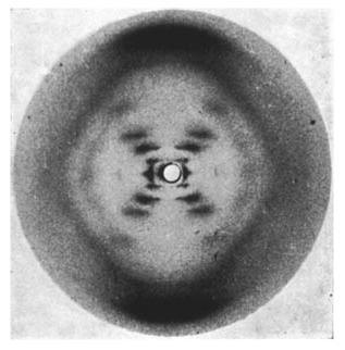
Sixty years ago this month Nature published the famous paper by Watson and Crick solving the structure of DNA. At the time many researchers pursued this goal, made difficult by the complexity of the DNA itself. A key contribution to the solution of the puzzle was the x-ray diffraction data provided by Rosalind Franklin. Indeed, without x-ray diffraction experiments this discovery would have been almost impossible at the time.

Rosalind Franklin’s x-ray diffraction image of DNA. (c) Nature Magazine. Franklin, R. & Gosling, R. G. Nature 171, 740-741 (1953) – doi:10.1038/171740a0
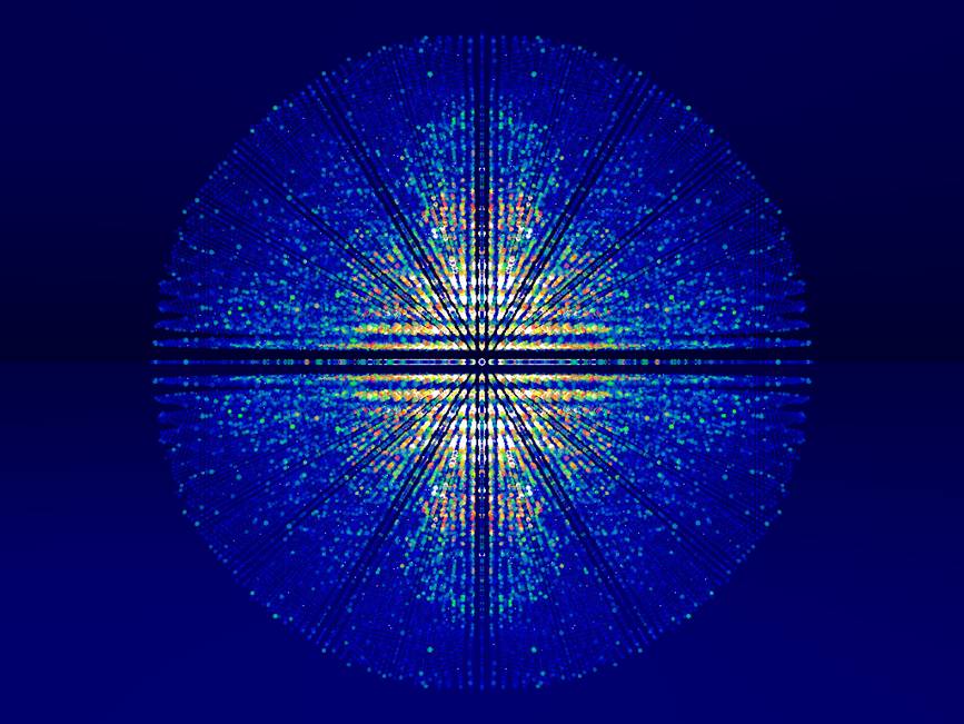
The way x-ray crystallography works is that a beam of x-rays is directed at a crystal, where the x-rays bounce off the atoms. Because the atoms in a crystal are periodically arranged, the x-rays form complex but regular patterns (such as the one seen for DNA). A detailed analysis of these patterns enables the precise determination of the crystal structure.
To this day such experiments aren’t easy. They require relatively large crystals and typically are done at major facilities such as electron synchrotrons. The synthesis of the crystals for these experiments can often be very difficult.
Yasuhide Inokuma, Makoto Fujita and colleagues from the University of Tokyo in Japan and the University of Jyväskylä in Finland have now developed a clever method that does away with many limitations of x-ray crystallography. Their method works with tiny amounts of material, only about a half to 5 micrograms are enough. This is around a millionth of a gram – truly tiny. The difference between a microgram and a gram is the same as that between a gram and a metric ton. In addition, another major advance of their method is that the target molecules don’t even need to be in a crystalline state. […]


April 1, 2013
1 Comment